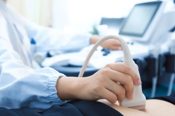
Ultrasound / Sonography

Ultrasound / Sonography
Diagnostic ultrasound, often known as sonography or diagnostic medical sonography, is a type of imaging that uses sound waves to create images of inside body structures. The photos can help doctors diagnose and treat a wide range of diseases and disorders. The majority of ultrasound exams are performed with an ultrasound device outside of your body, while some may require the placement of a tiny device inside your body. Ultrasound examinations are usually painless. An ultrasound examination typically takes 15 minutes to half an hour.
Here Are Some Services Include !
Elastography is a newer non-invasive medical imaging technique used to assess the stiffness of organs and other body structures. The most common application is to evaluate your liver fibrosis. New applications in breast, thyroid, prostate, kidneys and lymph nodes are emerging. Elastography uses low-frequency vibrations to deliver painless stimulation to the liver and other organs. The speed with which these vibrations flow through the organ is measured using ultrasound (US). This data is used by a computer to build a visual map of the stiffness (or elasticity). Elastography is useful in differentiating malignant from benign neoplasms (especially in breast), aiding in deciding the biopsy site more accurately, thus reducing negative biopsy rates.
Shear Wave Elastography (SWE) is a technique used in medical imaging to assess tissue stiffness, particularly in the context of liver fibrosis. A Shear Wave Fibrosis Scan is a specific application of SWE used to evaluate liver fibrosis non-invasively.
3D and 4D ultrasounds are advanced sonographic imaging features. It employs sound waves to create an images of your baby in your womb. A three-dimensional image of your kid is produced, whereas 4D ultrasounds produce a live video effect, similar to that of a movie, in which you may watch your baby grin or yawn. Ultrasounds in 3D and 4D have been shown to be safe in studies. Additionally, the photos might assist doctors in detecting and explaining a problem related to your foetus.
This Doppler uses a computer to convert sound waves into various colours. In real-time, these colours depict the pace and direction of blood flow. A newer sort of colour Doppler is the Power Doppler. It can show greater blood flow details than the traditional colour Doppler. A doctor may recommend an ultrasound to a pregnant woman when there is a need to closely monitor her baby's health and provide more useful advice and treatment. This method will assist a doctor in calculating a baby's growth at each stage
An echocardiogram creates images of your heart using sound waves. A doctor can watch your heart beating and pumping blood with this common test. This can be used by doctors to detect heart problems. It is essential for establishing the health of the heart muscle, particularly following a heart attack. It can also identify cardiac abnormalities or anomalies in unborn/ newborns. Echocardiography is a painless procedure. Only very rarely are there concerns with particular types of echocardiograms or when contrast is used during the echocardiogram.
A thoracic ultrasound, often known as a chest ultrasound, is a procedure that uses sound waves to produce detailed images of your chest. Ultrasound imaging creates images of your organs by using high-frequency sound waves and their echoes. Critical care ultrasonography (CCUS) includes thoracic ultrasonography, which allows the intensivist to assess the lung and pleural space. In the intensive care unit (ICU), it can reduce the use of routine chest radiography (CXR) and computerised tomography (CT). Thoracic ultrasonography is the "go-to" modality for imaging the lung and pleura in an efficient, cost-effective, and safe manner because of its ease of use, speed, repeatability, and dependability.
An abdominal ultrasound is a non-invasive procedure used to access the organs and structures within the abdomen. This includes the liver, gall bladder, bile ducts, pancreas, spleen, kidneys and pelvic organs.
A paediatric ultrasound produces images of the organs and soft tissues inside the body that may be viewed live on a computer screen using sound waves. It doesn’t use radiation and has no known harmful effects. It is very useful for evaluating the causes of abdominal, pelvic or scrotal pain in children.
A fetal echocardiogram (also known as a fetal echo) creates images of an unborn baby's heart using sound waves. This non-invasive ultrasound exam reveals the heart's structure and function. Fetal echocardiography is performed while you are lying down in a darkened room. It's similar to a standard pregnancy ultrasound.
In Obstetrics ultrasound, ultrasound waves are used to create images of a baby (embryo or foetus) inside a pregnant woman's uterus and ovaries. It is the recommended approach for monitoring pregnant women and their unborn babies because it does not involve ionising radiation and has no known side effects. One of the most important functions of obstetrical ultrasound is to confirm your pregnancy. They can be performed at any time during your pregnancy. Gynaecological ultrasound is used to diagnose many problems related to pelvic organs (Uterus and ovaries).
An anomaly scan, also known as a mid-pregnancy scan, is an ultrasound scan performed between the 18th and 21st weeks of pregnancy to examine the baby and the womb (uterus) more closely and determine where the placenta is located. The goal of the scan is to find any serious physical abnormality in the 2-dimensional (2-D) image of the baby in the womb.
Follicular monitoring is a series of scans or ultrasounds used to track the ovaries' follicle growth. Transvaginal ultrasound is used for follicular monitoring because it is more accurate (pelvis can be seen more clearly) and more convenient (you don't have to hold pee) than transabdominal ultrasound. A series of ultrasound vaginal scans are used to determine if a woman is ovulating and to pinpoint when a follicle ruptures and releases an egg. It's also known as follicle tracking, and it helps couples figure out when they should have intercourse for the maximum likelihood of conceiving.
Ultrasound imaging creates images of your interior organs by using high-frequency sound waves. Imaging scans can help doctors diagnose illnesses by detecting anomalies. A transvaginal ultrasound, also known as an endovaginal ultrasound, is a form of pelvic ultrasound that doctors use to evaluate all parts of the female reproductive system. The term "transvaginal" literally means "through the vaginal canal." This is a test that will only be conducted internally. Unlike a standard abdominal or pelvic ultrasound, in which the ultrasound wand (transducer) is placed on the exterior of the pelvis, this sonography includes your doctor or a technician inserting an ultrasound probe about 2 or 3 inches into your vaginal canal.
Tendonitis, bursitis, carpal tunnel syndrome, rotator cuff tears, joint issues, and abnormalities such as tumours or cysts are all disorders that can be diagnosed by musculoskeletal ultrasonography. The technology allows you to roam about during the treatment in addition to providing high-resolution photographs. Using the ultrasound's Doppler effect, which monitors moving substances, we can also focus on arteries and veins—and detect how blood is flowing.
Elastography is a newer non-invasive medical imaging technique used to assess the stiffness of organs and other body structures. The most common application is to evaluate your liver fibrosis. New applications in breast, thyroid, prostate, kidneys and lymph nodes are emerging. Elastography uses low-frequency vibrations to deliver painless stimulation to the liver and other organs. The speed with which these vibrations flow through the organ is measured using ultrasound (US). This data is used by a computer to build a visual map of the stiffness (or elasticity). Elastography is useful in differentiating malignant from benign neoplasms (especially in breast), aiding in deciding the biopsy site more accurately, thus reducing negative biopsy rates.
Why Choose us?
Choosing us over other, Navya Diagnostics have such world class facilities and doctors which you won’t get anywhere.
This hospital is not only a treatment center, in fact it’s a hope for many. We do our best in treating the patients. We know what the requirements of a patient are and to reduce extra burden, we keep working for their good health and quick recovery.
w
We gives you the best healthcare solutions.
If you have any query or want to share problems or any emergency, contact us without hesitation..
